The answer to Africa’s malaria challenge could fit in a pocket.
Texas A&M researchers hope to revolutionize the way the disease is identified by simply using a smartphone and a type of clip-on microscope. Their new technology was inspired by a study abroad project that exposed an A&M professor to the need for easily accessible malaria detection methods in Africa.
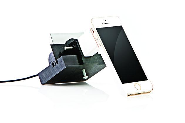
The small microscope can easily attach to a smartphone’s camera and a malaria diagnosis can be made anywhere. This device may make it possible for many more people to be tested and ultimately healed.
The softball-sized microscope holds a compact five-centimeter lens. The apparatus is attached to a phone case, which isn’t much different from regular cases. To use the microscope, the phone is simply inserted into the case.
Gerard Cote, A&M professor of biomedical engineering, is a lead designer on the project.
“I was part of a study abroad and was teaching some students in Rwanda, Africa,” Cote said. “As a part of their study abroad project, they had to come back with a medically related design idea, and I thought I would do the same.”
The malaria situation in Rwanda is improving, but it is still gravely serious. According to the Rwandan government, one in 13 children will not make it to the age of 5 due to this disease.
“While I was over there, I saw the need for malaria detectors because they have the medication, but they don’t know when to dole it out,” Cote said.
Standard testing for malaria is a long process in Rwanda that involves administering the test sending the test off to a central laboratory, and waiting for the results to get back.
“I thought if I could come up with a way of doing a mobile device that used a microscope apparatus, it could help a lot of folks over there so they could determine whether they had it or not and quickly receive the right medication,” Cote said.
Kokou Dogbevi is a biomedical engineering graduate student working on the second generation of the microscope.
“The first generation is used with the iPhone 5s,” Dogbevi said. “We 3D print the case for the phone, the lens space and everything.”
The current design uses the standard camera, but future versions of the device may work with a separate application.
“The goal is to develop an application to process the image that the phone receives but will be developed in the future,” Dogbevi said.
The technology in the device is simple. Malaria parasites are “birefringent” — when polarized light hits them, the vertically polarized light becomes elliptical and shows up as little spots on the camera. The microscope uses polarized light in a similar way that polarized sunglasses do to detect the disease.
Although the primary application for the device is malaria detection, it can also be used for a slew of other purposes.
“Anything that polarized microscopy is used for could potentially be looked at on the cell phone,” Pirnstill said.
One such thing that can be detected in this way is skin cancer.
“We are using the same concept to look at skin cancer,” Cote said. “The skin cancer cells show up much better under these polarized lights so it could be used for that as well.”
Casey Pirnstill, Class of 2012, was involved with the design and build of the phone microscope. He said the technology could produce better malaria detection due to its cost and portability.
“Malaria is so prominent and there really wasn’t a technology that can be used in the field,” Pirnstill said. “This device is portable and low cost.”


