


Texas A&M scientists are using cutting-edge physics to create real life medical breakthroughs.
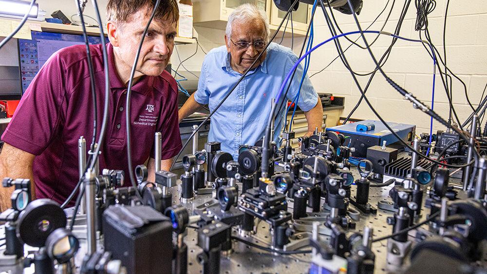
Professor of physics and biomedical engineering, Vladislav Yakovlev, Ph.D., and professor of physics and biological and agricultural engineering, Girish Agarwal, Ph.D., FRS, at A&M, along with Tian Li, Ph.D., assistant professor of physics at the University of Tennessee at Chattanooga, recently developed a new type of microscope that takes advantage of quantum physics in order to dramatically increase the resolution of images the microscope takes.
The microscopy technique the A&M team is investigating, Yakovlev said, relies on a process called Brillouin scattering, where a ray of light, such as a laser, is beamed into the sample being imaged. The light’s energy causes particles in the sample to vibrate, and the specific frequencies of the vibrations reveal the positions and identities of the particles. Once this is known, scientists can produce an image of the sample they are analyzing.
“Brillouin microscopy is a very special technique which can measure the mechanical properties of materials with very good special resolution, so we can see inside cells,” Yakovlev said. “For example, in cancer, cancer cells migrating from one side of the tissue to another or to a different tissue is driven by mechanical processes, so understanding these processes is important.”
The problem, Yakovlev said, comes from an interaction between light and living cells. When imaging anything, the light waves are subject to random interferences from the environment that cause the pictures to be blurrier. When it comes to Brillouin microscopy, which involves shining an intense light beam on a tiny sample, these typically small interferences can render images completely useless. To produce clearer images, the scientists want to use more powerful light beams to drown out the environmental noise. However, if the light beams are too powerful, scientists can risk damaging or killing cells when imaging them.
“When we image cells, we run into the problem where if we want to get more [clarity] we have to increase our light’s intensity. However, if we increase the intensity past a certain point we start causing damage,” Yakovlev said. “It seems like you can’t get both [image clarity and undamaged cells] at the same time just because you want to increase intensity to reduce your noise. But on the other hand, if you increase intensity, you damage your cells.”
In order to solve this, Yakovlev said his team has built a device that takes advantage of a physical phenomenon known as entanglement. In the minuscule world of quantum physics, a group of particles, in this case light waves, can be generated in a manner so they effectively act as a single entity, and actions that affect one wave will affect the others. Entanglement forms the basis for many cutting-edge technologies, such as quantum computing, and may be the key to birds’ ability to circumnavigate the globe, a phenomenon noted by M. Suhail Zubairy, Ph.D., professor of physics at A&M, in his book “Quantum Mechanics for Beginners.”
“[A] highly counterintuitive feature of quantum mechanics is that two or more objects can form an ‘entangled’ state,” Zubairy writes. “[Intuitively], if we have two objects such as two balls that are placed a large distance apart, they are strictly independent of each other. No matter what we do to one, it cannot influence the other. Quantum mechanically we can form states for the two objects such that, even when the two objects are very far from each other, what we do to one object can influence the state of the other.”
For Yakovlev and his team, its application lies in removing noise from microscopy images. Yakovlev said the new device his team has developed creates two entangled beams of light. When interference affects one wave, the effects are reflected in the entangled wave as well. The device compares the two waves and finds extraneous interference patterns by analyzing the similarities in the waves. Once this is known, the device removes the interference from both waves, resulting in an image that is significantly clearer than an image produced by standard Brillouin microscopy.
“We are essentially making clever manipulation of light beams so we know that there’s noise present in both beams and we know that these noises correlate,” Yakovlev said. “We know both these laser beams are entangled and so we know that the noise is going to be the same for both of them, so we can just find out the similarities and subtract them, and that’s how we make that clear image at the end.”
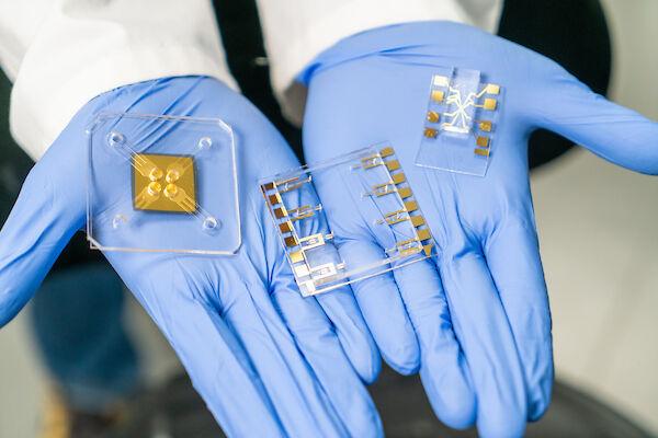
Li, at the time a graduate student at the University of Maryland, was hired by A&M to build the laboratory for the device. Li said the device has successfully created microscopic images of several biological samples while also measuring their composition.
“The whole lab was built from the ground up. The lab is relatively new but we have the capability of generating quantum entangled light [for the device],” Li said. “We have already [imaged] cancer cells. We’ve also tried the brain of the fruit fly.”
Marlan Scully, Ph.D., professor of physics and director of the Institute for Quantum Science & Engineering at A&M, sponsored the research and provided the laboratory space. Scully said learning more about the applications and interactions between quantum physics and biology is a high priority for A&M.
“Combining what we know now about quantum mechanics and biology, we come to a new field called quantum biology,” Scully said. “That’s the essence of what people like Agarwal and Yakovlev are trying to understand. It’s a big function of my lab at [the Institute for Quantum Science and Engineering].”
In a press release published by A&M’s college of engineering, Agarwal said the new device represented a breakthrough for medical imaging technology as well as the growing development of real-world applications for quantum physics.
“It’s a new milestone in the capabilities of Brillouin microscopy and imaging extensively used for biosystems,” Agarwal said in the release. “It becomes part of an international effort to develop quantum sensors for diverse applications like brain imaging, [and] biomolecule structure mapping”